Overview
What is calcific tendonitis?
Calcific tendonitis is a build-up of calcium deposits within one or more of the four tendons that meet at the top of the shoulder joint. Together these four tendons are known as the rotator cuff. The build-up of calcium deposits leads to pressure and chemical irritation in the tendon. This can be extremely painful.
The calcium deposits can also rub or irritate a small, fluid filled sac (called a bursa), located between the bone on the top of the shoulder (called an acromion), and the rotator cuff tendons. The bursa is designed to enable the smooth gliding of the rotator cuff under the acromion. When irritated or inflamed it is less able to do this and movement results in pain.
In many cases, the calcific tendonitis can lead to symptoms of subacromial impingement. This happens when the calcium deposits cause rotator cuff tendons to be pinched against the acromion
Why does it happen?
We still don't know for sure why calcific tendonitis affects some people and not others. It can affect all age groups and a wide range of people who take part in a wide range of different activities.
How common is it?
Although any of the rotator cuff tendons may suffer from calcific tendonitis, it is the supraspinatus tendon that is most commonly affected. Age can play a role, as calcium deposits tend to be more common in the 30 to 50 age group. It may be related to some activities or occupations
Symptoms
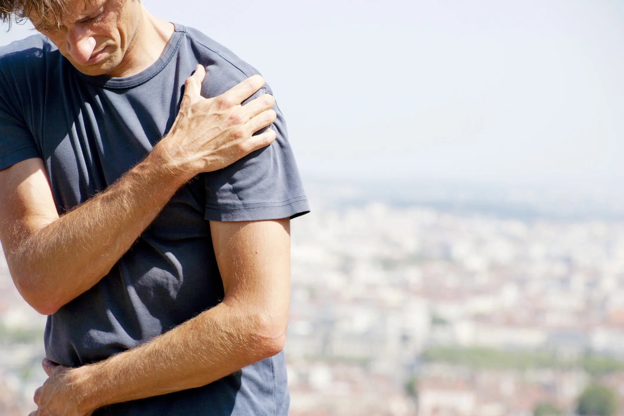
The most common symptom is pain. In addition, as calcific tendonitis can cause impingement in the shoulder joint, it can also present many of the same symptoms as subacromial impingement. This may include a painful arc of movement when the arm is lifted out to the side and up to your ear. Some people report significant pain at night, which can interrupt their sleep. Others often describe a "locking" sensation in their arm when attempting certain movements.
It is important to report any symptoms of neck, shoulder, upper arm or hand pain to Mr. Cole. In addition, remember to tell him if you feel pins and needles or tingling in your arm or hand. This is because they may indicate that the pain is coming from your neck, via the nerves in your arm.
Investigating the problem
Physical examination
Mr. Cole will talk to you about your shoulder symptoms. He will examine your shoulder and look at areas such as your range of movement, whether your shoulder is tender to the touch and whether it makes any sound when moved. He will use some specific clinical tests and test the rotator cuff tendons

X-RAY
An x-ray can provide an excellent "picture" of bones and joints and can clearly show evidence of calcific deposits.
Ultrasound
An ultrasound uses high frequency sound waves to produce images of the body. The procedure is painless and is particularly good at providing images of any calcific build up from angles that would be harder to achieve with an an x-ray.
Treatment options
Non-surgical treatment
Painkillers
Pain medication and anti-inflammatory medicines will help to ease your symptoms of pain and stiffness. If your symptoms are mild, this may be enough to keep your condition manageable.
Physiotherapy
An individual programme of exercises will be devised for you. These may include exercises to strengthen the muscles around your shoulder blade, improve your posture, strengthen your rotator cuff and improve stretching. Although the exercises may at times be hard work, tight or uncomfortable, they should not be painful. Should you need any other form of treatment, such as injections or surgery; physiotherapy will form an important part of your rehabilitation and recovery.
Injections
Depending on the extent and duration of your symptoms, you may benefit from a steroid injection. Steroids (such as cortisone) are good at reducing the inflammation. However, it is unlikely that you will able to have more than three steroid injections during one year.
Ultrasound-guided barbotage
An ultrasound image is used to identify the exact area of calcification. Under ultrasound guidance and with local anaesthetic the calcific deposit can be punctured, possibly aspirated and flushed out. A steroid injection is also applied to treat the inflammation. Barbotage is completed as an outpatient, day procedure. You may be in some discomfort following it, so it is a good idea to ask someone to drive you home. You should be able to return to driving the following day though.
The procedure is successful in 60-70% of patients, although some people require two treatments. If you do not respond to the barbotage, you may need surgery, although Mr. Cole will advise you of this.
Extracorporeal Shock Wave Treatment

Using a specialised machine shockwaves are passed through the tendon. This is done usually without the need for local anaesthetic or injections. This can be very effective in some patients and 3 treatments as an outpatient are usually required. It has recently been studied and approved for calcific tendonitis in the shoulder by NICE (National Institute for Clinical Excellence)
Exercises
Exercising your shoulder is an important part of your recovery. It can help to minimise stiffness and avoid further possible complications such as muscle atrophy (where your muscles start to waste away because they are not used). An exercise programme will be devised for you by your team of physiotherapists, in conjunction with Mr. Cole. It will last for several weeks and is something you will need to do every day.
Surgical treatment
Should you need surgery, Mr. Cole will advise you on what type of treatment would best suit you and your situation. He will choose the most appropriate and least invasive for you.
Arthroscopic treatment
This is a keyhole surgery where a camera is used to help identify and target specific problem areas. A Subacromial Decompression is performed and any large calcific deposits can be removed from the tendon at the same time. This is normally completed as day surgery and is minimally invasive.
Exercises
Following your surgery, your team of physiotherapists will work with you to devise an exercise programme. This will be designed with advice fromMr. Cole to help you recover as quickly and efficiently as possible.
Surgery
About arthroscopic excision of calcific tendonitis
The need to have surgery for the treatment of calcific tendonitis is not common. The vast majority of people who develop the condition may be successfully treated with steroid injections, physiotherapy or ultrasound
guided barbotage and occasionally shock wave therapy.
However if the symptoms are ongoing and painful, you may need surgery to increase the amount of space between the flat bone at the top of your shoulder (acromion) and tendons of your shoulder joint (rotator cuff). This procedure is called subacromial decompression and will prevent the painful pinching you have been experiencing.
If you have a build-up of calcium in the area, Mr. Cole will clean this away (debride it) in a procedure called arthroscopic excision of calcific tendonitis, at the same time that the arthroscopic subacromial decompression is being performed. This is the least invasive type of surgery and will leave minimal scarring.
Preparing for your operation
Your arthroscopic excision of calcific tendonitis operation will be day surgery. This means that you won't normally have to stay overnight in hospital. However, you may feel more comfortable if you bring your own dressing gown, slippers and toiletries.
You will be having a general anaesthetic. This can make you feel woozy for a little time after the operation, so you will need to arrange for someone to take you home. It is important that you don't eat or drink anything for six hours prior to your admission into hospital, although you may drink water up to two hours prior to admission.
If you normally wear make-up or nail varnish, please remove it prior to your admission. You will also need to bring all prescribed medicines and supplements, in their original containers, with you to the hospital.
Understanding your operation
After admission, you will be seen by Mr. Cole and by your specialist anaesthetist. They will talk about the operation and the anaesthetic with you and, where possible, discuss your preferences. Nothing will be done without your permission on the day.
Anaesthetic and pain relief
The operation is carried out under a general anaesthetic. After your admission, you will be given a pre-med. This is usually in tablet or liquid form; It will help you relax before your surgery.
You will then be taken to the anaesthetic room, where you will be given the anaesthetic. This may either be a gas to breathe or an injection. This will be all you remember about the operation, as you will fall asleep at this point and awake after the operation in the recovery room.
Usually you will be given an interscalene nerve block during the operation. This acts as an excellent pain relief and for a short while after your operation your shoulder and arm may feel numb. When this wears off, your shoulder can feel more sore. You will be given some painkillers to take after the operation. When you begin to feel sensation returning to your shoulder (often a "pins and needles" feeling), you should start taking the pain medication that you have been given.
Don't wait for your shoulder to start hurting, as pain is best managed before it gets acute. To keep the pain under control, use your medication regularly to begin with. After a couple of days, you can begin to lower the amount you take and then cease the medication altogether once any pain has subsided. If the pain does not seem to get better, or if you need more pain medication, please contact Mr. Cole.
A cold compress can also help with pain relief and swelling at the site of your operation. If you use an ice pack it is important you keep the wounds dry.
Risks
All operations involve a small element of risk. In arthroscopic excision of calcific tendonitis, these can include:
Complications relating to the anaesthetic such as sickness, nausea or rarely cardiac, respiratory or neurological issues (less than 1% each, i.e. less than one person out of one hundred).
Infection. These are usually superficial wound problems. Very rarely deep infection may occur many months after the operation (less than 1%).
Persistent pain or stiffness in or around the shoulder (5-10% of patients could still have some symptoms after the operation).
Damage to the nerves and blood vessels around the shoulder (less than 1%).
The need to re-do the surgery is rare. In less than 5% of cases, further surgery is needed within 10 years.
Please discuss these issues with Mr. Cole if you would like further information.
Recovery
Follow-up appointments
You will usually be invited to attend an outpatient clinic within 2 weeks of your operation, where the wound will be examined and your dressing removed. After about 6-12 weeks you will be asked to return so that Mr. Cole can check on your progress. You may discuss any concerns you have during these appointments.
Alternatively, should you have a concern, you may telephone Mr. Cole's clinic at any time following your operation.
The wound
Arthroscopic excision of calcific tendonitis is completed by keyhole surgery. This means you will only have two to three small puncture wounds. You will not have any stitches, only small sticking plaster strips. Keep the wounds dry until they are healed, which is normally within five to seven days. You can wash or shower and use ice packs, but protect the wounds with waterproof dressings. These will be given to you on your discharge from hospital. Avoid using spray deodorant, talcum powder or perfumes near or on the wounds until they are well healed. The dressing will normally be removed at your first follow-up appointment.
The sling
You will be given a sling to wear immediately following the operation. This is for your comfort and can be discarded within a few days. You can take the sling on and off as you wish, although you might find it more comfortable to wear your sling at night.
Sleeping
Sleeping may be uncomfortable for a while. It's best to avoid sleeping on the side of your operation. If you choose to lie on the other side, you can rest your arm on pillows placed in front of you. A pillow placed behind your back can help prevent you from rolling onto your operated shoulder during the night. If you are lying on your back to sleep you may find placing a thin pillow or small rolled towel under your upper arm or elbow will enhance your comfort.
Your recovery
Different people recover at different rates. However, by about three weeks following your operation you should find that movement below shoulder height becomes more comfortable. You should also be able to move your arm into most positions, including above shoulder height, although this might still be a little painful.
By three months you should feel a lot better. However, it can take from six to nine months to fully recover. You will continue to improve for up to a year following the procedure.
Driving
You may begin driving one week after your operation or when you feel comfortable. Check you can manage all the controls and it is advisable to start with short journeys.
Returning to work
The best time for you to return to work depends on how you feel following the operation and on the type of work you do. If your job is largely sedentary with minimal arm movements close to your body, you may be able to return within a week. Most people are able to return to work within a month at most. However, if you have a heavy lifting job or one with sustained overhead arm movements you may require a longer period of rehabilitation. It is best to discuss this with Mr. Cole and with your physiotherapy team.
Returning to sport and leisure activities
Your ability to start these activities will be dependent on pain, range of movement and strength that you have in your shoulder. It is wise to avoid sustained or powerful overhead movements (such as trimming a hedge, some DIY, racket sports, front crawl) for a few months. These types of movement will put stress on the subacromial area and may take longer to become comfortable. It is best to start with short sessions involving little effort and then gradually increase the effort or time for the activity. Your physiotherapy team will be able to give advice tailored to you and your situation.
Physiotherapy
You will be shown exercises by the physiotherapist and you will need to continue with the exercises once you go home. They aim to stop your shoulder getting stiff and to strengthen the muscles around your shoulder. We have outlined these early exercises here. Your physiotherapy team will also devise a longer term programme tailored for you and your situation.
Use pain-killers, ice packs or both to reduce any pain before you begin exercise, if necessary. It is better to do short frequent sessions of physiotherapy several times a day, rather than one long session. Aim to exercise for five to ten minutes, four times a day.
It is normal for you to feel aching, discomfort or stretching sensations when doing these exercises. However, intense or lasting pain (such as pain that lasts for more than 30 minutes) is an indication to change the exercise by doing it less forcefully or less often.
Post operative exercises
Phase 1 Exercises
You can begin these exercises immediately after your operation.
1. Pendulum (shown for left shoulder)
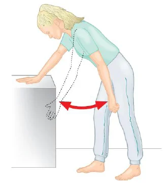
Lean forwards with support
Let your arm hang down
Swing arm
1. forwards and back
2. side to side
3. around in circles (both ways)Repeat 5–10 times each movement
2. Lower trapezius (shoulder blade exercises) (shown for both shoulders)
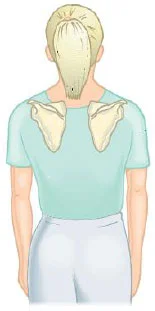
Do this exercise either sitting or standing
Keep your arms relaxed
Square your shoulder blades (pull them back and slightly down)
Do not let your back arch
Do not let elbows move backwards (you can clasp your hands in front of you, to discourage this!)
Hold for 10 seconds
Repeat 10 times, “little and often” during the day
3. Twisting outwards (external rotation) (shown for right shoulder)
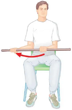
Sit holding a walking stick, or rolling pin, or umbrella
Keep your elbow into your side throughout
Push with your unaffected arm, so that the hand of your problem side is moving away from the mid-line (you can also do this exercise lying down)
Do not let your body twist round to compensate
Repeat 5–10 times
4. Arm overhead (flexion in lying) (shown for left shoulder)
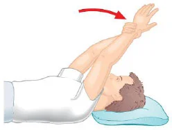
Lie on your back on your bed or the floor
Support the arm of your operated shoulder with your other hand at the wrist and lift it up overhead
Do not let your back arch
Try to get your arm back towards the pillow or floor
You can start with elbows bent
Repeat 5–10 times
5. Arm overhead (flexion in standing) (shown for right shoulder)
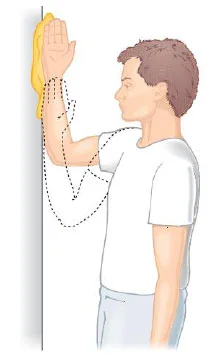
Stand facing a wall with the elbow of your operated shoulder bent and your hand resting against the wall
Slide your hand up the wall, aiming to get a full stretch. (If necessary, use a paper towel between your hand and the wall to make it easier)
Repeat 10 times
6. Shoulder blade exercise (shown for right shoulder)
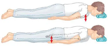
Lie on your front, with your head face down supported by a rolled-up towel under your forehead, or with your head turned towards your operated shoulder
Keep your arms relaxed by your side
Lift your shoulder straight up in the air. Try and keep a gap approximately 5cms between the front of your shoulder and the bed or floor you are lying on
Hold your shoulder up for 30 seconds
Repeat 4 times
You may progress this exercise by lifting your arm up and down (elbow straight) while keeping the shoulder blade up all the time
I would like to thank Professor Carr and Jane Moser of the Oxford Shoulder and Elbow Clinic for allowing us to reproduce some of this text and illustrations from their patient information.